Plantar fasciitis is one of the most commonly treated foot conditions and one of the most frustrating. Stretching, orthotics, corticosteroid injections, and rest produce temporary relief for many patients without producing lasting recovery. One possible reason in chronic cases is that treatment is being directed at the endpoint of the problem rather than at what is loading it.
Part of that picture is well established in the published literature. Part of it is a clinical hypothesis that has not been tested. This article separates the two, because the difference matters for what a patient should reasonably expect.
What the Evidence Establishes: The Calf
The link between calf tightness and heel pain is not a fringe position. It is reflected in how foot and ankle surgeons already operate.
A study quantifying the relationship found a strong statistically significant correlation between gastrocnemius tightness and the severity of heel pain in patients with plantar fasciitis. A current concepts review of plantar fasciopathy notes that gastrocnemius tightness has been associated with ankle dorsiflexion stiffness and plantar fascia injury, and that isolated proximal medial gastrocnemius release has been reported for cases that have not responded to other treatment. That is surgery on the calf to resolve pain in the heel.
A systematic review of factors associated with chronic plantar heel pain found evidence of an association with decreased ankle dorsiflexion, alongside body mass index in non-athletic populations, the presence of a calcaneal spur, increased age, decreased first metatarsophalangeal joint extension, and prolonged standing. Reduced ankle dorsiflexion is, functionally, a tight calf.
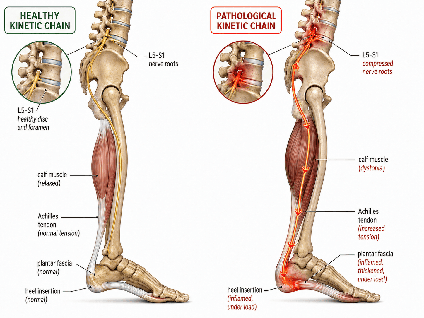
The mechanical logic is straightforward. A shortened calf acts as a pulley under constant load, transmitting tension through the Achilles tendon and concentrating force on the plantar fascia at its attachment on the heel bone. The fascia develops inflammation and microtearing at that attachment, producing the characteristic pain on first steps in the morning. Treat the fascia while the calf remains short and the force generator stays active.
This also explains why Achilles tendinopathy and plantar fasciitis frequently occur together. Both sit at the ends of the same tensioned structure. Finding one is a reason to examine the other.
Where the Hypothesis Begins: The Spine
If a tight calf loads the plantar fascia, the next question is what is tightening the calf. This is where the neuromyofascial framework makes a proposal that the published literature does not yet address.
The lower limb functions as a continuous mechanical chain. Nerve roots exiting the lumbar and sacral spine travel through the hip, thigh, calf, and into the foot. The hypothesis is that where those nerve roots are compressed or irritated, whether from disc pathology, fibrosis, or neuromyofascial scarring at the lumbar or sacral levels, the effects would travel the length of the chain.
Specifically, the proposal is that nerve root involvement at L5 or S1 creates motor neuropathy in the muscles those roots supply, particularly the calf. The gastrocnemius and soleus would respond with dystonia, a state of chronic involuntary tension. A dystonic calf would then produce exactly the mechanical picture the published research describes: sustained tension through the Achilles and abnormal load on the plantar fascia.
That upstream link has not been demonstrated. Searches of the indexed literature do not turn up studies establishing a lumbar or sacral contribution to plantar fasciitis. What exists is a documented calf-to-fascia relationship and an anatomically plausible but untested account of what may drive the calf dysfunction in some patients. Presenting it as anything more settled than that would misrepresent the evidence.
The Layers of a Complex Presentation
Simple presentations may involve only the calf and foot, and often respond to local rehabilitation directed at both.
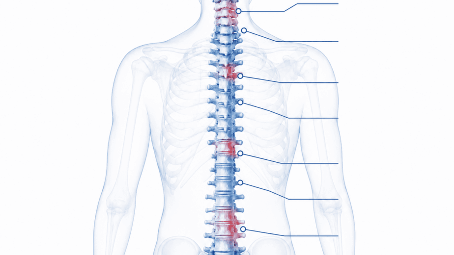
Chronic and more complex presentations, in my clinical experience, tend to involve several regions at once. The injury map in these cases typically spans the lower lumbar spine or sacrum, the pelvic and hip musculature, the hamstrings, the calf, and the foot. Each region may contain areas of dystonia or fibrosis contributing to the tension loading the plantar fascia.
Hip musculature is frequently overlooked. The piriformis, gluteus medius, and related pelvic muscles, when chronically tight or scarred, alter lower limb alignment and loading mechanics through the whole leg. That altered loading would compound whatever is happening further up and further down the chain. This is a clinical observation rather than a documented mechanism.
When the Foot May Not Be the Right Place to Look
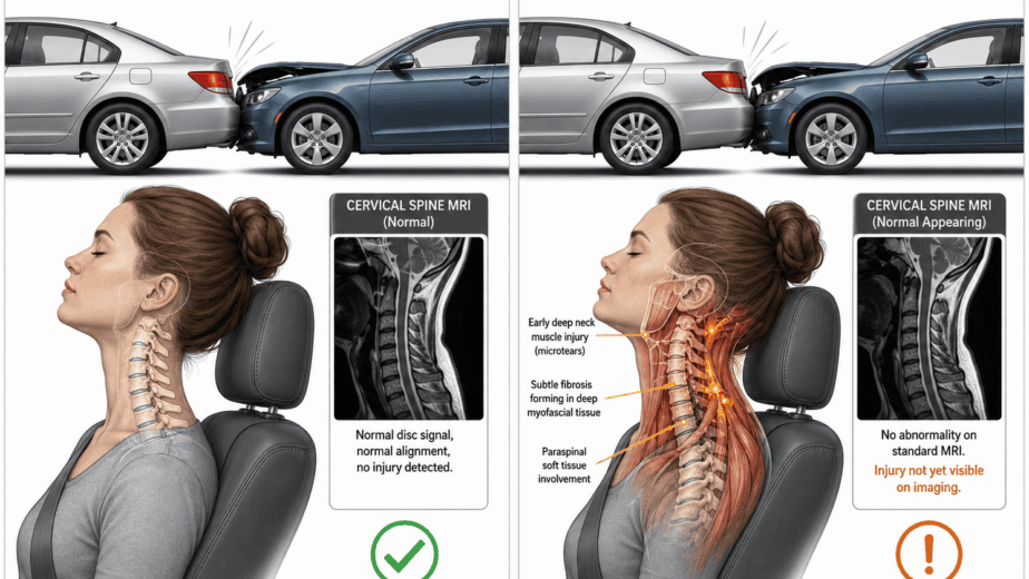
In a small number of cases, heel pain has persisted through exhaustive local treatment, thorough lumbar and sacral assessment, and hip rehabilitation. In some of those patients, neuromyofascial assessment has identified findings in the upper thoracic or cervical spine.
That may seem counterintuitive, since the cervical spine and the plantar fascia are anatomically distant. The reasoning behind investigating it is that the spinal cord runs continuously from the brainstem downward, and that dysfunction at the level of the cord itself can in principle generate motor abnormalities in limbs well removed from the site of involvement. Whether that mechanism operates in plantar fasciitis specifically is an open question, and the observation that these findings are sometimes present does not establish that they are causing the heel pain.
These are unusual presentations. Most plantar fasciitis does not warrant cervical spine investigation. In the genuinely treatment-resistant patient who has worked through every local and regional approach without result, widening the assessment is reasonable.
What Assessment Should Cover
A thorough neuromyofascial assessment of chronic plantar fasciitis maps potential contributing sites from the lumbar and sacral spine down through the hip, pelvis, hamstring, calf, and foot, with the aim of identifying which appear to be active contributors and how they may be loading the fascia.
Care is then directed at the sites the map identifies rather than applied as a generic protocol. Whether that approach produces better outcomes than standard care in a controlled comparison has not been tested, and that study would be worth running. What can be said is that the calf is a documented contributor, that ankle dorsiflexion is a recognized associated factor, and that assessment stopping at the fascia is assessment that stops early.
Plantar fasciitis that keeps coming back may not be a stubborn foot problem. It may be an incomplete investigation.
The information in this article is educational and informational in nature. It is not intended as a substitute for professional medical advice, diagnosis, or treatment. If you are experiencing chronic foot pain or plantar fasciitis that has not responded to standard treatment, consult with a qualified healthcare provider to discuss the options appropriate for your situation.