Carpal tunnel syndrome is one of the most commonly diagnosed and surgically treated conditions in upper limb medicine. The standard model is straightforward: the median nerve is compressed within the carpal tunnel at the wrist, producing numbness, tingling, and weakness in the thumb, index, middle, and part of the ring finger. Decompress the tunnel and the symptoms resolve.
For many patients that model works. For a significant proportion who do not fully recover or who eventually relapse, it may be incomplete, and one possible reason is that the wrist was not the only place the nerve was in trouble.
The Nerve Runs from the Neck to the Hand
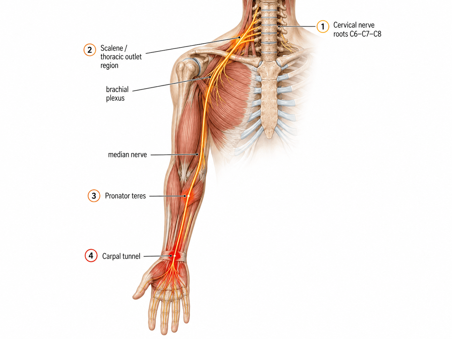
The median nerve does not begin at the wrist. It originates from nerve roots in the lower cervical spine, primarily C6, C7, and C8, and travels through the brachial plexus, across the shoulder, down the forearm, and into the hand. Any point along that pathway where the nerve is compressed, tethered, or subjected to chronic traction is a potential contributor to median nerve symptoms in the hand.
This is the concept behind what the literature calls double crush syndrome: the proposal that a nerve under proximal compression becomes more vulnerable to symptomatic injury at a distal site. It was described by Upton and McComas in The Lancet in 1973. Their electromyographic study of 115 patients with carpal tunnel syndrome or ulnar nerve lesions at the elbow found electrophysiological evidence, often supported by clinical symptoms, of associated neural lesions in the neck in 81 cases, roughly 70 percent. They argued the association was not coincidental but reflected serial constraints on axoplasmic flow along the nerve fibre.
It is important to say plainly that the double crush hypothesis is contested and has been since it was proposed. Critics have pointed out that in most of Upton and McComas’s cases there was no direct evidence the cervical pathology was actually affecting the nerve roots, and that the authors suggested a relationship between neck and wrist symptoms without demonstrating one. The hypothesis has been widely cited and widely challenged.
What has held up better than the causal claim is the observation of co-occurrence. A cross-sectional study of 291 patients with electromyographically confirmed cervical radiculopathy found carpal tunnel syndrome in 62.2 percent of them. The same study found no correlation between the severity of the radiculopathy and the severity of the carpal tunnel syndrome at any cervical level, and the authors concluded this argues against a causal relationship between the two.
That is a fair summary of where the evidence sits. These conditions occur together far more often than chance would predict. Whether the proximal problem causes the distal one is not established. The clinical implication does not depend on settling that question: if both are frequently present, assessing both is reasonable, and assessing only one is a decision to leave something unexamined.
Where Else the Median Nerve Can Be Compromised
Beyond the cervical spine, the median nerve passes several places where it can be affected before it reaches the wrist, and this is better documented than the cervical question.
A 2022 review by Löppönen and colleagues in the Journal of Clinical Medicine addressed proximal median nerve compression directly. Their conclusion is that these compressions should be understood as a spectrum of mild to severe lesions along a branching nerve, producing variable symptoms, rather than as the neat categories of pronator syndrome and anterior interosseous nerve syndrome that the literature has traditionally used. They note that clear objective findings are not always present, and that diagnosis therefore depends on a thorough understanding of anatomy and careful clinical testing rather than on tests alone.
That last point explains a great deal. A review of pronator syndrome in the Journal of Hand Surgery notes that its symptoms often overlap with carpal tunnel syndrome and that electrodiagnostic studies are frequently negative, which makes correct diagnosis challenging. A proximal contribution that produces no abnormality on nerve conduction testing is a proximal contribution that will not appear in a standard workup.
Thoracic outlet involvement belongs in the same discussion. Compression or tethering of the brachial plexus as it passes between the scalene muscles in the neck and beneath the pectoralis minor in the chest wall can produce upper limb pain, numbness, tingling, and weakness that resembles distal entrapment.
The Neuromyofascial Proposal
What the neuromyofascial framework adds to this picture is a proposed mechanism linking those sites, and it should be read as a hypothesis rather than an established sequence.
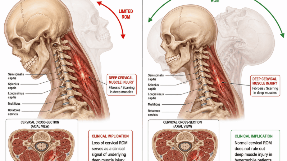
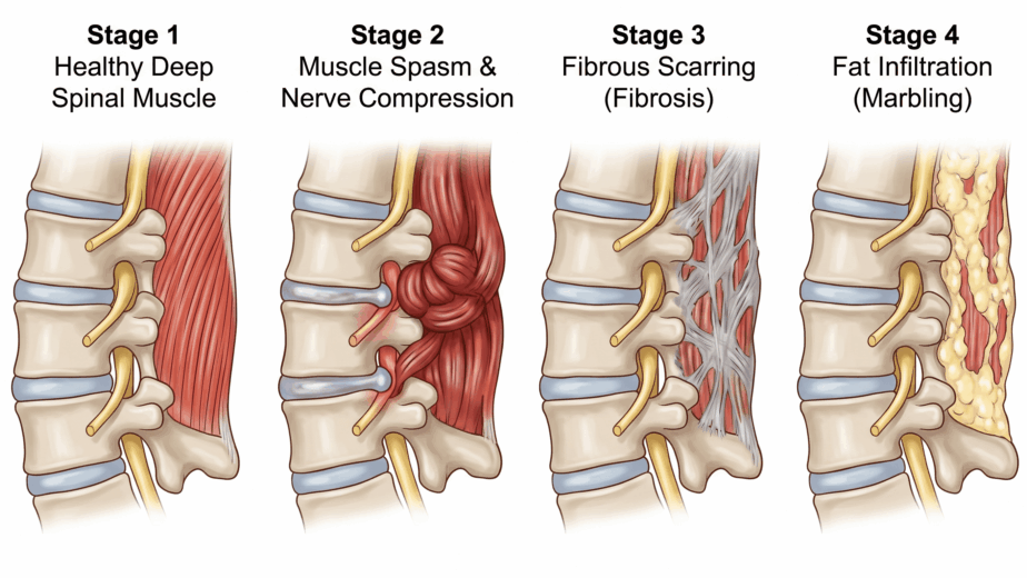
The proposal is that the injury sequence often begins in the cervical spine, whether from a whiplash event, repeated neck strain from prolonged desk work, or cumulative upper back injury. Neuromyofascial scar tissue accumulates in the lower cervical region, frequently without appearing on standard MRI, and irritates the nerve roots supplying the arm.
That irritation would produce a motor neuropathy: partial impairment of the motor signal traveling down the arm. Muscles supplied by those roots, including neck and shoulder girdle muscles, the scalenes, and eventually the forearm muscles, would lose normal tone regulation, progressively shorten, and develop dystonia. As the scalenes and pectoralis minor tighten, they would create compression and tethering of the brachial plexus. Further down, tightness in the pronator muscles of the forearm would add compression points along the median nerve before it reaches the wrist.
By the time the nerve arrives at the carpal tunnel in this model, it is under chronic traction from several points above and is less mobile than it should be. The carpal tunnel becomes the last pinch point on an already loaded nerve. Releasing that pinch without addressing what has been loading the nerve from above would produce relief that fades as the traction mechanism continues.
None of that chain has been demonstrated experimentally. Each link is individually plausible and several are documented in isolation. The sequence as a whole is a clinical hypothesis that would require prospective study to test.
Ulnar Neuritis Follows Similar Logic
The ulnar nerve, which supplies the little finger and ring finger and originates from C7 through T1, follows a parallel course through the same cervical and thoracic outlet territory. Ulnar neuritis, in which the nerve is entrapped at the elbow, was part of the original Upton and McComas cohort and shows the same pattern of frequent proximal findings.
In the neuromyofascial model, ulnar presentations often involve contractures and trigger points in the latissimus dorsi and triceps that may create traction on the nerve as it travels toward the elbow. That is a clinical observation rather than a documented mechanism, and it is offered as a reason to examine the whole limb rather than as an explanation of the condition.
When Surgery Is and Is Not the Answer
Surgical carpal tunnel release remains appropriate and necessary where compression at the wrist is severe, where there is significant motor loss or muscle wasting, or where the condition is acute and progressive. The literature does not support delaying surgery in those presentations, and nothing in this article should be read as a reason to.
What the literature does support is that median nerve symptoms can arise from more than one place along the nerve, that proximal contributions often produce no abnormality on standard testing, and that a proportion of patients continue to have symptoms after wrist decompression. In those cases, examining the cervical spine, thoracic outlet, shoulder girdle, and forearm is a reasonable next step rather than a fringe one.
The neuromyofascial approach is to map those potential contributors and address them alongside local care. Whether doing so improves outcomes compared with standard management has not been tested in a controlled study, and that trial would be worth running.
The wrist is where the symptoms are. In some patients, it is not the only place worth looking.
The information in this article is educational and informational in nature. It is not intended as a substitute for professional medical advice, diagnosis, or treatment. If you are experiencing symptoms of carpal tunnel syndrome or upper limb nerve pain, consult with a qualified healthcare provider to discuss the diagnostic and treatment options appropriate for your situation.