For nearly 30 years, I have been pioneering the field of neuromyofascial science, focusing particularly on the challenge of diagnosing and treating invisible spinal injuries—a realm where traditional medical imaging often falls short. My research into fat water indexing has opened new doors in understanding how injuries transform spinal muscle tissue, leading to chronic pain conditions that elude conventional treatment methods. This introduction is an invitation to explore the groundbreaking implications of this research, which not only offers a more accurate diagnosis of spinal injuries but also provides a predictive model for recovery, promising a new era in the management of neuromyofascial pain.
It is common in medical practice to see patients who have undergone a whiplash event, and complain of chronic spinal pain, yet have normal or near normal reports of their spine whether it be Xray, CT scan or even an MRI.
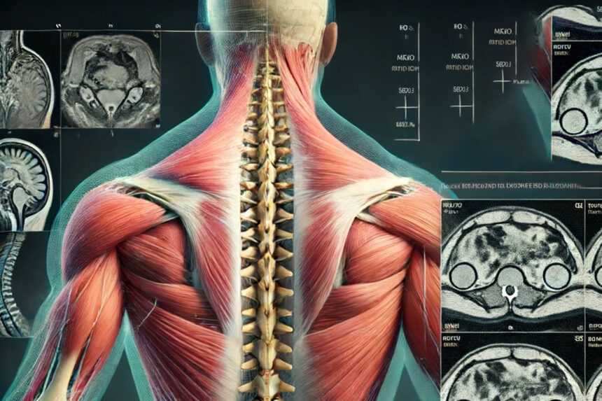
Our imaging whether it be x ray, ultrasound, CT scan or MRI has great difficulty in detecting significant changes in the spinal soft tissue that can cause changes in our normal spinal anatomy and ultimately cause catastrophic failure in our spinal segments.
There have been some recent changes in newer MRI technology reporting and discussing some soft tissue injuries, and similar reporting is now present in some ultrasound of the spine, but it is still very limited.
However, the newer soft tissue spinal MRI research has been progressing for over a decade.
I am following several groups of researchers in the US and Australia appreciating what I call the “not so soft” tissue around the spine which develops post whiplash.
The MRI analysis is named fat water indexing, and I will explain what it really implies.
Fat water indexing in a spinal MRI application looks at how the deep spinal muscles become injured and the muscle is replaced with fat over time. The fat develops over about 3 months after a whiplash event, as the fat replaces the scarred damaged deep spinal muscle tissue.
The idea is that we cannot yet see the injury or specific changes in the web of muscles around the spine, but we can see the formation of fat in the muscle; and we can call it marbling, like steak.
James Elliott, a leading spinal MRI researcher looking at whiplash injuries in the neck has shown repeatedly in multiple studies where infiltrates of fat formed over time AFTER injury, usually seen as early as 2 weeks after the MVA, and the study could predict who would recover with rehabilitation, and who would never recover based on the MRI of the neck at around the 2-4 week period simply by measuring the muscle replacement with fat by percentage. At three months you could really see the fat marbling in the spinal muscle, and link it fairly directly to certain types of chronic pain, non recovery, and even the development of anxiety and PTSD.
In multiple medical studies using FWI MRI of the spine, we see that when the fat content exceeds 20% in the deep spinal muscles, persistent pain will occur, and the patient will not recover with traditional rehab care.
Other radiology research groups lead by Mengiardi showed similar changes in the lower back whereby intrinsic muscles of the spine had higher concentration of fat in the specific areas of the lower back whereby chronic pain was reported by the patients when compared with asymptomatic volunteers.
In this study though , spinal fat was present in all chronic low back pain patients, whether from a specific injury or from repetitive wear and tear or aging which tells us that intrinsic spinal muscle scarring with fat replacement may predict most spinal pain problems.
I personally think this is true, and I have formulated much of my research around intrinsic spinal pathology.
But further research will be required.
Finally, Pfirrmann, another MRI research group leader, showed similar changes in rotator cuff muscles as far back as 2004, In this study the fat development was predictive of the degree of tearing of the rotator muscle, so by looking at fat content you can risk assess a tear in a muscle; so Fat water indexing is not really that new, just not appreciated by medical science , ….yet.
Moreover, I explain the effect as like in the spine, that the rotator muscles are denervated of motor signal from the neck, which causes the supraspinatus to shorten and scar replacing the muscle with fat allowing for the muscle to become more easily torn.
This suggests that rotator muscle tears are preceded by spinal nerve signal loss, which could be seen by spine FWI MRI, and is nicely consistent with my neuromyofascial pain theory and science.
So, to summarize, the formation of fat or marbling in muscle after injury has very significant implications in both the spine and the limbs, and in prediction of chronic pain, non recovery, tearing of muscle and probable denervation of the muscles leading to other areas of pain.
I think this info is important in managing and recovering people in car accidents.
Reflecting on the advancements in neuromyofascial science, particularly the application of fat water indexing to spinal injuries, it’s evident that we are on the cusp of a major breakthrough in understanding and treating conditions that have long challenged the medical community. The ability to visualize the transformation of muscle to fat post-injury not only offers a deeper insight into the nature of chronic pain but also heralds a new approach to rehabilitation and recovery. As we continue to refine these techniques and integrate them into clinical practice, the promise of a more effective, personalized treatment for spinal and neuromyofascial conditions becomes increasingly tangible. My commitment to this field is driven by a profound belief in the potential to improve the lives of those affected by such injuries, underscoring the importance of innovation, empathy, and patient-centered care in the journey toward healing.
Medical Disclaimer:
The information provided in this article is for educational and informational purposes only and is not intended as a substitute for professional medical advice, diagnosis, or treatment. Always seek the advice of your physician or another qualified health provider with any questions you may have regarding a medical condition or treatment and before undertaking a new health care regimen, regardless of your location. Never disregard professional medical advice or delay in seeking it because of something you have read on this website.
AI Disclaimer:
The images and abstracts featured in our blog posts are generated using artificial intelligence. While we strive for accuracy and relevance, there may be occasional discrepancies or errors. We appreciate your understanding and encourage readers to consider the context and intent behind these AI-generated elements.