Most people assume that when a muscle is injured, it either heals or it does not. What they rarely hear is that under certain conditions, injured muscle appears to change into something else.
Over three decades of spinal research and clinical work, I have studied what happens to the deep intrinsic muscles of the spine following injury, particularly after whiplash. Part of what follows is documented in the imaging literature. Part of it is a clinical model of how the process unfolds, which the research has not yet tested. The distinction matters, so I have marked it throughout.
What the Imaging Research Documents
The most solid ground here is the fat and water MRI work led by James Elliott and colleagues, which measures fat content within the deep cervical muscles directly.
In one prospective study, 36 patients were imaged within one week of whiplash injury, then at two weeks and three months. There was no difference in muscle fat infiltration between groups at enrolment. By two weeks, patients who would go on to report severe disability at three months already showed significantly higher fat infiltration in the cervical multifidus than those who recovered. A threshold of 20.5 percent fat infiltration at two weeks predicted poor outcome at three months with 87.5 percent sensitivity and 92.9 percent specificity. The authors concluded that muscle degeneration occurs soon after injury, but only in those patients with poor functional recovery, and that routine imaging protocols may need to be reconsidered.
A larger study of 97 patients presenting to an emergency department after a motor vehicle collision followed them to twelve months. Neck muscle fat infiltration measured at one week, together with a traumatic distress score, significantly predicted neck disability at twelve months. Patients who recovered had lower fat infiltration than those with mild or moderate to severe outcomes at every time point measured.
A confirmatory study found the infiltration concentrated in the medial portions of the deep cervical extensors, with the magnitude significantly greater in patients with severe chronic symptoms at twelve months.
So the phenomenon is real, it is measurable within weeks, it tracks strongly with who does badly, and it is concentrated in exactly the deep stabilizing muscles this discussion concerns.
What the Research Does Not Settle
Two findings complicate any simple causal story, and both come from the same body of work.
The authors of the 97-patient study state plainly that it is unclear whether higher fat infiltration represents a pre-existing phenotype or a result of the trauma. If some patients arrive at their collision with deep spinal muscle already in a more vulnerable state, then fat infiltration measured a week later may be partly a marker of who they were before the accident rather than a product of it.
The second is more interesting still. An earlier study following 44 patients found that initial post-traumatic stress symptom severity mediated the relationship between pain intensity and muscle fat infiltration at six months, while loss of cervical range of motion did not. A psychological factor sat in the middle of the chain between pain and tissue change.
Neither finding undermines the importance of the tissue change. Both mean that a purely mechanical account of how it arises is incomplete.
The Clinical Model
What follows is my working account of the sequence. It is consistent with the imaging findings but has not been tested as a causal chain, and should be read that way.
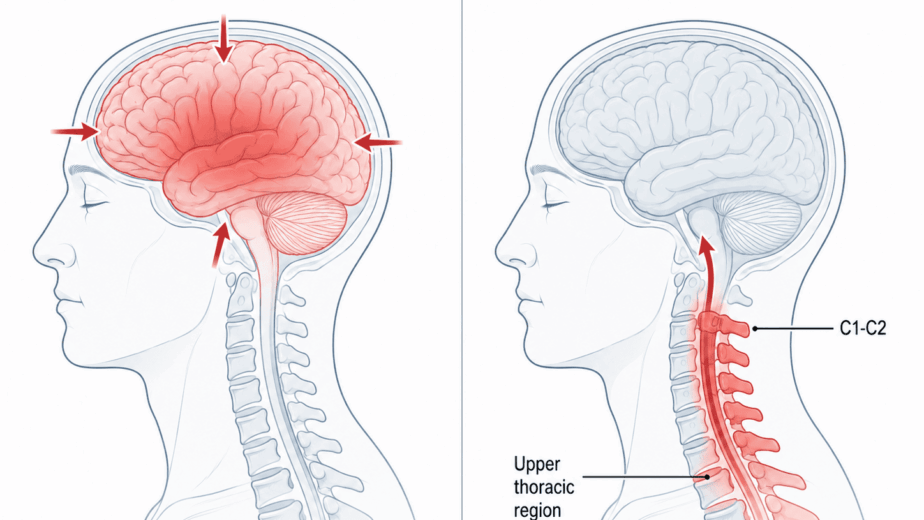
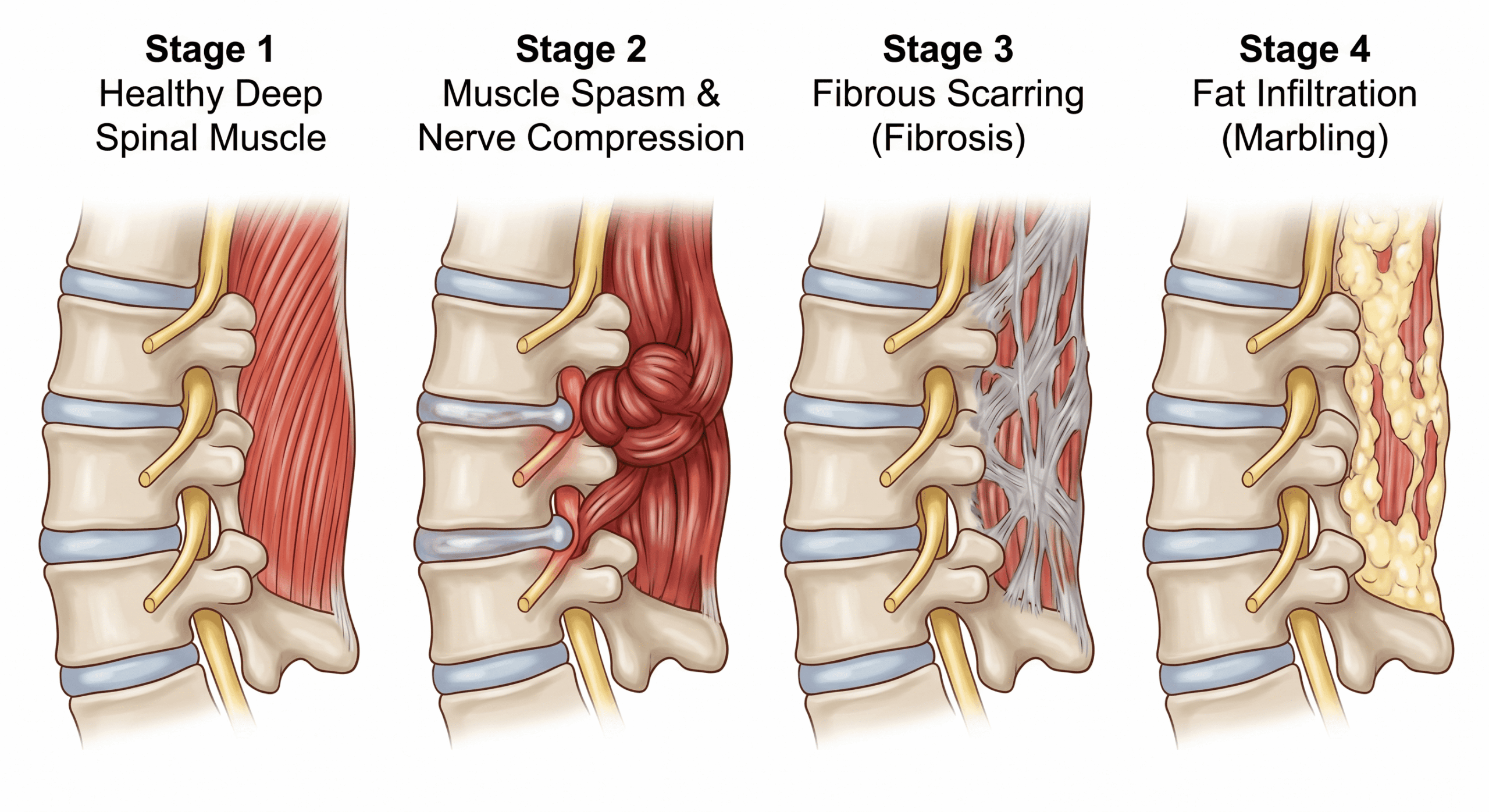
A whiplash event does not simply strain the neck. It may directly injure the deep intrinsic spinal muscles, the small stabilizing muscles running alongside and between vertebrae. Those muscles respond with spasm, which is normally protective. But because they sit in close proximity to nerve roots, discs, and bony structures, persistent spasm may begin compressing those structures.
My proposal is that this compression affects the dorsal rami, the nerve branches supplying the deep spinal muscles, and that reduced nerve signal disrupts normal motor function such that the muscle loses its ability to relax. The result would be dystonia: persistent involuntary shortening that does not resolve on its own.
Once established, the cycle would become self-reinforcing. Sustained spasm generates scarring within the muscle over subsequent weeks and months. Dense fibrous change begins to immobilize spinal segments, which compounds compression on discs and nerve roots and alters spinal mechanics further. Muscle deprived of normal signal and locked in a state it cannot escape progressively stops functioning as contractile tissue, and is replaced by fat. That endpoint is the part the imaging research measures directly.
The imaging confirms the destination. Whether the route I have described is how patients get there is a hypothesis that would require dedicated study, including electrophysiological assessment of the dorsal rami, to test properly.
Why Standard Rehabilitation Can Reach a Ceiling
Physiotherapy, exercise, and manual therapy are valuable for many spinal pain presentations. They work when the underlying tissue can respond to load, movement, and progressive strengthening.
Where tissue has been substantially replaced by fat and fibrous change, the picture is different. This is not a failure of rehabilitation as a discipline, and it is not an argument against trying it. It is a question about what the tissue being asked to respond is actually made of.
It is worth being honest about the limits of what is known here. I am not aware of studies establishing whether deep cervical fat infiltration is reversible, or whether any particular intervention reduces it. The research establishes that it predicts poor outcome. Whether changing it changes the outcome is a separate question, and one nobody has yet answered. That gap is a reason for research rather than a reason for confidence in any approach, including mine.
What This Means for Patients
Chronic spinal pain after an injury is often accompanied by confusion and frustration. Imaging comes back normal or near normal. Rehabilitation produces partial improvement that then plateaus. Symptoms persist despite reasonable effort.
Part of what may be missing is recognition that measurable structural change can occur in the deep spinal tissues within weeks of injury, that standard imaging protocols are not designed to detect it, and that it predicts who will still be symptomatic a year later. A patient whose routine MRI is unremarkable has not been shown to have normal deep cervical muscle. They have been shown to have an unremarkable routine MRI.
The neuromyofascial approach is to assess these tissues directly, stage what is found, and direct care accordingly rather than managing symptoms at the surface. Whether that produces better outcomes than existing rehabilitation in a controlled comparison has not been tested. The imaging research makes a strong case that something structural is happening early in the patients who do not recover, and that identifying it sooner would be worth doing.
The information in this article is educational and informational in nature. It is not intended as a substitute for professional medical advice, diagnosis, or treatment. If you are experiencing chronic spinal pain following an injury, consult with a qualified healthcare provider to discuss the options appropriate for your situation.