One of the most consistent observations in thirty years of clinical practice is that patients with chronic pain often do not know where their pain is coming from. They know where it lands. They know where it hurts. But the site where they feel the symptom and the site that may be generating it are frequently not the same place.
This gap between symptom location and injury origin is one of the central problems in chronic pain medicine. Treating the location of pain rather than a contributing source may be part of why so many patients improve temporarily and then plateau, or why a new symptom appears somewhere unexpected after an old one settles.
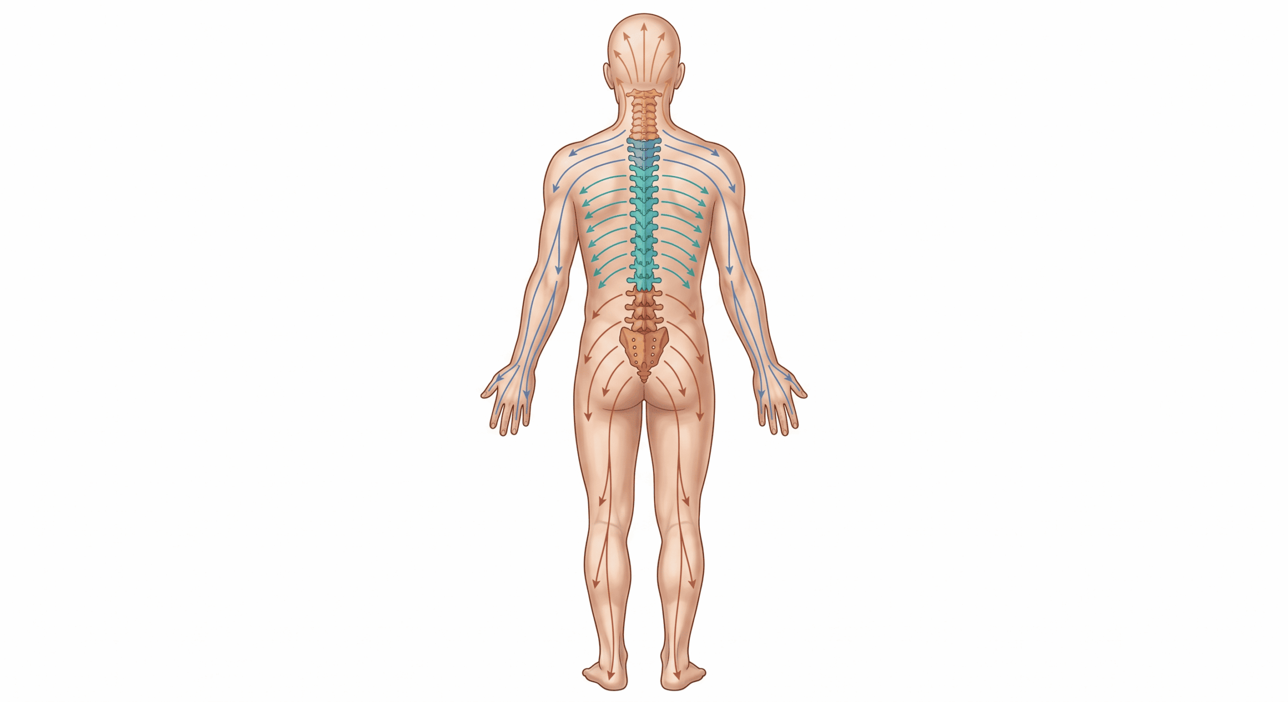
What follows is a working map: a framework for how different regions of the spine may generate different symptom patterns. Some of it is well established in the published literature. Some of it is clinical observation that has not been formally studied. I have tried to be clear throughout about which is which.
The Upper Neck and Craniocervical Junction
I divide the cervical spine into upper and lower regions because they generate distinctly different symptom patterns.
The upper neck and craniocervical junction, meaning the region from the base of the skull down through C1 and C2, is the most neurologically complex area of the entire spine. Injury here tends to produce craniofacial and sensory symptoms: migraine-type headaches, facial pain, balance disturbance and vertigo, tinnitus, visual difficulty, and light or sound sensitivity.
The anatomy behind this is established rather than speculative. Nociceptive afferents from the first three cervical spinal nerves converge with trigeminal afferents in the trigeminocervical nucleus, in the upper cervical spinal cord. Because of that convergence, pain from structures supplied by C1 to C3 can refer into trigeminal territory, meaning the head and face, and pain from trigeminal territory can refer back into the cervico-occipital region. The literature places the proportion of chronic unilateral headache that is cervicogenic at roughly 15 to 20 percent.
The dizziness connection has its own literature. Cervicogenic dizziness is a recognized condition with published diagnostic criteria. The cervical spine carries densely developed proprioceptive receptors whose input is integrated with visual and vestibular information in the central nervous system. When cervical proprioceptive input changes and conflicts with those other inputs, dizziness can result. A systematic review and meta-analysis of 13 randomized trials involving 898 patients found moderate-quality evidence that manual therapy reduces cervicogenic dizziness along with cervical spine and balance symptoms, with a stronger effect when combined with exercise therapy, though the authors rated the evidence for the combination as very low quality.
Tinnitus follows a similar pattern. Somatosensory tinnitus is a recognized subtype in which input from the cervical spine and temporomandibular area alters the tinnitus perception. A screening tool developed from 7,981 survey responses identifies it using four criteria, including whether tinnitus and neck or jaw pain rise and fall together, tension in the suboccipital muscles, and whether the tinnitus changes with somatic manoeuvres. That model detects somatosensory tinnitus with 82 percent accuracy.
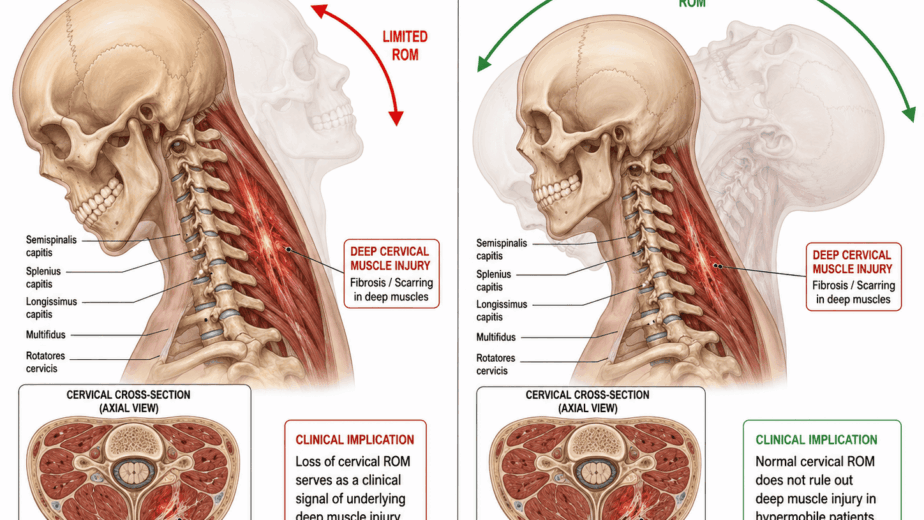
The craniocervical junction is also the transition point where the spinal cord becomes the brainstem. The neuromyofascial proposal is that injury and fibrosis here may tether the spinal cord from below, transmitting upward tension into the brainstem and cranial nerves. That specific mechanism is a hypothesis rather than a demonstrated finding. What is established is that this region refers symptoms into the head, face, ears, and balance system through documented pathways, which is why upper cervical injury so often produces presentations mistaken for primary brain or inner ear pathology.
The Lower Neck
Lower cervical spine injuries, from approximately C3 through C7 and into the upper thoracic spine, tend to generate a different pattern. The classic presentation is tension-type headache: a band-like pressure across the front and sides of the head, distinct from the more severe and often unilateral pain associated with upper cervical injury.
Lower neck injury also affects the upper limbs. Numbness, tingling, and weakness in the arms and hands are common. Carpal tunnel syndrome and ulnar neuritis, which produce different distributions of hand and finger numbness, may in some patients involve a cervical contribution rather than isolated wrist or elbow entrapment alone.
The evidence there needs stating carefully, because it cuts both ways. A cross-sectional study of 291 patients with electromyographically confirmed cervical radiculopathy found carpal tunnel syndrome in 62.2 percent of them, a high rate of co-occurrence. The same study found no correlation between the severity of the cervical radiculopathy and the severity of the carpal tunnel syndrome at any level, and the authors concluded this argues against a causal relationship. The two conditions travel together often. Whether one drives the other is unsettled. The defensible clinical position is that both warrant assessment rather than that either explains the other.
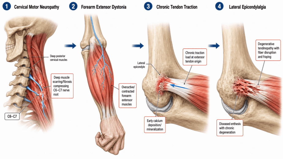
A pattern I observe frequently, which I refer to as myofascial thoracic outlet syndrome, belongs here as a proposed clinical entity rather than a recognized diagnosis. In this presentation, muscles of the neck and shoulder develop dystonia and fibrosis creating tethering around the brachial plexus. The result is diffuse global arm numbness rather than the distribution-specific numbness of carpal tunnel or ulnar neuritis. Tennis elbow, golfer’s elbow, hand and thumb pain, and grip weakness may also be downstream presentations. This is a clinical observation framework and has not been formally studied.
The Thoracic Spine
The thoracic spine is the most underinvestigated region of the spine in standard practice. In motor vehicle accidents it absorbs a significant portion of the force but is rarely assessed with the thoroughness applied to the cervical or lumbar regions. Part of the reason is practical, since thoracic injuries are difficult to visualize and quantify on standard imaging. Part is historical, since clinical attention has concentrated on the neck and lower back.
In my clinical experience the thoracic spine matters considerably in complex whiplash and chronic pain presentations. It is prone to accelerated kyphosis, an exaggerated forward curve, and to retrolisthesis, a form of vertebral slippage creating mid-back instability. Both may contribute to chest pain, rib pain, painful breathing, and gastrointestinal symptoms including reflux and bowel irregularity.
I also propose that spinal cord tethering can develop quietly in this region and contribute to symptoms elsewhere. A patient with treatment-resistant cervical pain may have a contributor in the thoracic spine that is not producing local upper back pain. A patient with lower limb neurological symptoms may have a thoracic component a lumbar-focused workup will not find. These are clinical observations offered as a reason to widen the assessment, not as established mechanisms.
I regard the thoracic spine as the structural foundation of both the cervical and lumbar regions. The neck and lumbar spine emerge from it. How it is positioned, how it moves, and where it is injured affects how both regions above and below it function.
The Thoracolumbar Junction
The transition point between the thoracic and lumbar spine, roughly T10 through L1, deserves its own section, and here the published literature is ahead of where most clinical practice sits.
Maigne Syndrome, also called thoracolumbar junction syndrome, was described by Robert Maigne in the late 1980s. It is characterized by pain at the iliac crest and tenderness on palpation at the junction between the lower thoracic and upper lumbar vertebrae. The critical feature for this discussion is where the pain presents. The disorder affects nerves at the thoracolumbar junction, but the symptoms typically appear in the lower abdomen, around the pelvic region, and in the groin. Any spinal joint between T9 and L2 can develop lesions and nerve impingement capable of producing referred pain, and patients generally respond well to manual techniques.
A review of the condition describes it as an often unrecognized and treatable cause of low back pain, with two variants: a central form arising from facet joint changes at the thoracolumbar junction, and a peripheral form involving impingement of the medial branch of the superior cluneal nerve. That review lists pseudosciatica and pseudo-visceral pain among its keywords, which is a precise description of the problem this whole article is about. Pain that appears to come from the sciatic distribution or from an internal organ, arising from a spinal segment well away from where it is felt.
My own clinical observation adds to this picture. The thoracolumbar junction commonly takes load in significant acceleration-deceleration events. When it does, the iliopsoas muscle, which attaches near this region and runs through the pelvis into the hip, may go into spasm, twisting the lumbar spine and producing the pelvic asymmetry and apparent leg length discrepancy that manual practitioners frequently identify. I also associate this region with hip and groin pain, hip joint degeneration, constipation, bladder dysfunction, and difficulty fully straightening the spine. The iliopsoas mechanism is my proposal. The referral pattern itself is documented.
The Lower Lumbar and Sacral Spine
The L1 through L4 zone primarily affects the front, side, and inner thigh. Quadriceps weakness, adductor pain, and hip flexor dysfunction are common presentations of nerve root compromise here, and are readily misattributed to hip joint pathology or groin strain.
The L4 through S4 zone generates the familiar patterns of sciatica: pain, numbness, tingling, or weakness in the back of the legs, calves, and feet.
The sacral region deserves specific mention because it is sometimes dismissed on the basis that there are no intervertebral discs at sacral level. That reasoning overlooks the possibility that spinal fascia in the sacral canal may constrict or tether nerve roots even without disc material, producing leg and foot symptoms a disc-focused workup would not identify. This is a proposed mechanism rather than a documented one, and it is the least evidenced claim in this article. I include it because the clinical pattern recurs, not because the mechanism has been demonstrated.
Reading the Map
This is a general framework, not a complete picture. Spinal injuries do not respect boundaries. A patient with significant whiplash rarely injures only one region. Upper, mid, and lower back injuries commonly coexist and interact, producing a broader symptom picture than any single region would alone.
The value of the map is not as a lookup table from symptom to spinal level. It is in the principle that symptoms have anatomical contributors, and that those contributors are sometimes located at a distance from where the pain is felt. That principle is not a neuromyofascial invention. Trigeminocervical convergence, cervicogenic dizziness, somatosensory tinnitus, and Maigne syndrome are all documented examples of exactly this, each with its own literature, and each routinely underrecognized in practice.
Specialist assessment of the symptomatic site is appropriate and often sufficient. An audiologist examining the ear in tinnitus, a surgeon examining the wrist in carpal tunnel syndrome, and a podiatrist examining the foot in plantar fasciitis are each doing the right thing for most patients. The map suggests an additional starting point for the cases that do not resolve: trace the pattern back toward the spine and check whether something upstream is contributing. In chronic and treatment-resistant presentations, that tracing is worth doing.
The information in this article is educational and informational in nature. It is not intended as a substitute for professional medical advice, diagnosis, or treatment. If you are experiencing chronic pain that has not responded to standard treatment, consult with a qualified healthcare provider to discuss the options appropriate for your situation.