Skip to contentSkip to sidebarSkip to footer
Neuromyofascial Science
Menu Close
  • HOME
  • CONDITIONS
    • MS
    • Fibromyalgia
    • Post-Concussion
    • TMJ & Tinnitus
    • Migraines
    • Elbow Pain
    • Foot Pain
    • Sciatica
  • PATIENTS
    • Browse All Conditions
    • Patient Success Stories
    • Latest Videos
  • CLINICAL RESOURCES
  • COLLABORATE
  • ABOUT
    • NMF Science Education
    • TNPC Treatment Info
    • NMF team
Close
  • HOME
  • CONDITIONS
    • MS
    • Fibromyalgia
    • Post-Concussion
    • TMJ & Tinnitus
    • Migraines
    • Elbow Pain
    • Foot Pain
    • Sciatica
  • PATIENTS
    • Browse All Conditions
    • Patient Success Stories
    • Latest Videos
  • CLINICAL RESOURCES
  • COLLABORATE
  • ABOUT
    • NMF Science Education
    • TNPC Treatment Info
    • NMF team
Neuromyofascial Science
  • Home
    • Hospital
      Doctor
      Medical Center
      Private Clinic
      Medical Lab
      Dentist
  • Pages
    • About Us
    • Our Mission
    • Services
    • Departments
    • Our Doctors
    • FAQ
    • Patient Success Stories
    • Pricing
    • Locations
    • 404 Page
    • Service Plus
    • AI Features
  • Services
    • About Us

      Learn about our hospital and values

      Services

      Explore our wide range of medical services

      Departments

      Discover our specialized medical departments

      Blog

      Get the latest health news and tips

      Contacts

      Contact us for appointments or inquiries

      Testimonials

      Read patient stories and feedback

  • Blog
    • Blog – Standard
    • Blog – Grid
  • Contact Us
Four-stage medical illustration showing the sequence from cervical motor neuropathy to lateral epicondylalgia. Stage 1 shows deep muscle scarring compressing C6-C7 nerve roots in the cervical spine. Stage 2 shows overactive contracted forearm extensor muscles from impaired nerve signal. Stage 3 shows chronic traction load at the extensor tendon origin with early calcium deposition. Stage 4 shows degenerative tendinopathy with fiber disruption and diseased enthesis.
Conditions, Performance and Sport

Why Athletes Keep Getting Re-Injured: The Spinal Origin of Tendinopathy

4 days ago0Comments
Professional sports medicine has access to extraordinary resources. The best imaging available. Expert physiotherapists, surgeons, and rehabilitation specialists. Nutritional and biomechanical support at every level.…
Split anatomical illustration showing normal airway versus post-whiplash airway in lateral cervical spine cross-section. Left panel shows open circular airway lumen with healthy pharyngeal muscles and labeled cervical vertebrae C1-C7. Right panel shows C3-C5 nerve disruption with amber highlight, dystonic pharyngeal muscles, and significantly narrowed oval airway lumen with amber highlight indicating obstruction.
Conditions, NMF Science Explained

When Whiplash Disrupts Sleep: The Cervical Spine and Sleep-Disordered Breathing

4 days ago0Comments
Sleep disruption is one of the most commonly reported but least investigated consequences of whiplash injury. Patients describe difficulty falling asleep, frequent nighttime waking, unrefreshing…
Posterior anatomical illustration of the human body showing the spine divided into four color-coded zones. Amber upper cervical zone with referral arrows toward the head, blue lower cervical zone with arrows toward the shoulders and arms, teal thoracic zone with arrows across the mid-back, and red lumbar zone with arrows sweeping into the hips and legs.
Conditions, NMF Science Explained

A Map of the Spine: How Spinal Injuries Generate Symptoms From Head to Foot

4 days ago0Comments
One of the most consistent observations in thirty years of clinical practice is that patients with chronic pain often do not know where their pain…
Anatomical illustration of the right arm and upper torso showing the median nerve pathway from cervical nerve roots C6-C7-C8 through four numbered tethering points: cervical nerve roots, scalene and thoracic outlet region, pronator teres in the forearm, and the carpal tunnel at the wrist, with progressive orange-to-red coloring indicating cumulative nerve vulnerability.
Conditions, NMF Science Explained

Carpal Tunnel Syndrome: Why the Wrist Is Usually Not Where It Starts

4 days ago0Comments
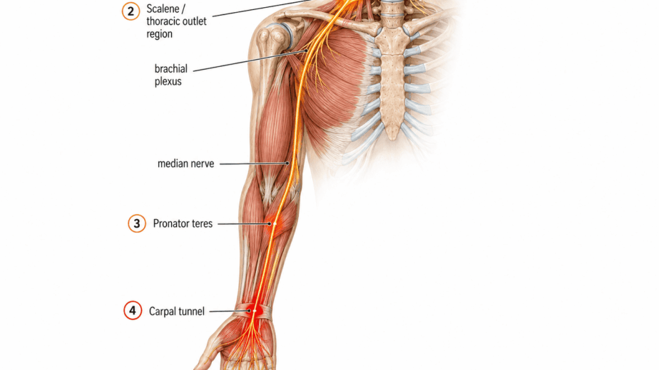
Carpal tunnel syndrome is one of the most commonly diagnosed and surgically treated conditions in upper limb medicine. The standard model is straightforward: the median…
Conditions, NMF Science Explained

Jaw Pain and Ringing in the Ears: Why the Neck Is Often the Missing Piece

4 days ago0Comments
Jaw pain and ringing in the ears are treated as separate problems. One gets sent to a dentist or oral surgeon. The other goes to…
Search
Categories
  • Conditions
  • NMF Science Explained
  • Injury and Recovery
  • Performance and Sport
  • Research and Clinical Insights
Recent Posts
Super Contractures: The Invisible Aftermath of Spinal Injury
Injury and Recovery, NMF Science Explained
Super Contractures: The Invisible Aftermath of Spinal Injury
June 9, 2026
Why Athletes Keep Getting Re-Injured: The Spinal Origin of Tendinopathy
Conditions, Performance and Sport
Why Athletes Keep Getting Re-Injured: The Spinal Origin of Tendinopathy
June 9, 2026
Neuromyofascial Science

Neuromyofascial Science is a precision-based clinical framework that maps the specific anatomical sources of chronic pain, neurological dysfunction, and structural pathology. Developed by Dr. G. Blair Lamb over 30 years of clinical innovation, NMFS uses a comprehensive neuromyofascial audit to build a patient-specific map of injury patterns and structural drivers, giving patients and their care teams a more precise picture of what is actually driving their condition.

Links
  • Home
  • Conditions
  • Patient Stories
  • Clinical Resources
  • Collaborate
Get in Touch
X-twitter Youtube Instagram Linkedin Tiktok

© 2026. All Rights Reserved.

Site by Neuromyofascial Science