Jaw pain and ringing in the ears are treated as separate problems. One goes to a dentist or oral surgeon. The other goes to an audiologist. Both usually receive treatment directed at the symptom site, and both frequently fail to fully resolve.
What the separate specialist model can miss is that these two conditions share anatomy. In a subset of patients, neither the jaw nor the ear is where the problem is being generated. The cervical spine is.
Somatosensory Tinnitus Is a Recognized Condition
Tinnitus originating in the inner ear involves cochlear damage or auditory nerve dysfunction and responds to audiological approaches. But a substantial portion of chronic tinnitus can be altered by what the neck and jaw are doing, and this is established rather than speculative.
Somatosensory tinnitus, sometimes called somatic tinnitus, is defined as tinnitus in which changes in somatosensory input from the cervical spine or temporomandibular area alter the tinnitus perception. It has diagnostic criteria. A screening tool developed from 7,981 survey responses identifies it using four questions: whether tinnitus and neck or jaw pain increase and decrease together, whether there is tension in the suboccipital muscles, whether the tinnitus can be modulated somatically, and whether the patient has bruxism. That four-item model detects somatosensory tinnitus with 82 percent accuracy and 82.5 percent sensitivity.
The clinical confirmation is straightforward and can be done in an examination room. If jaw clenching, head rotation, or pressure on specific neck muscles changes the pitch or volume of the ringing in real time, the tinnitus has a somatic component. That single observation determines whether treatment directed at the neck has any plausible route to affecting the sound.
The Trigeminal Cervical Complex
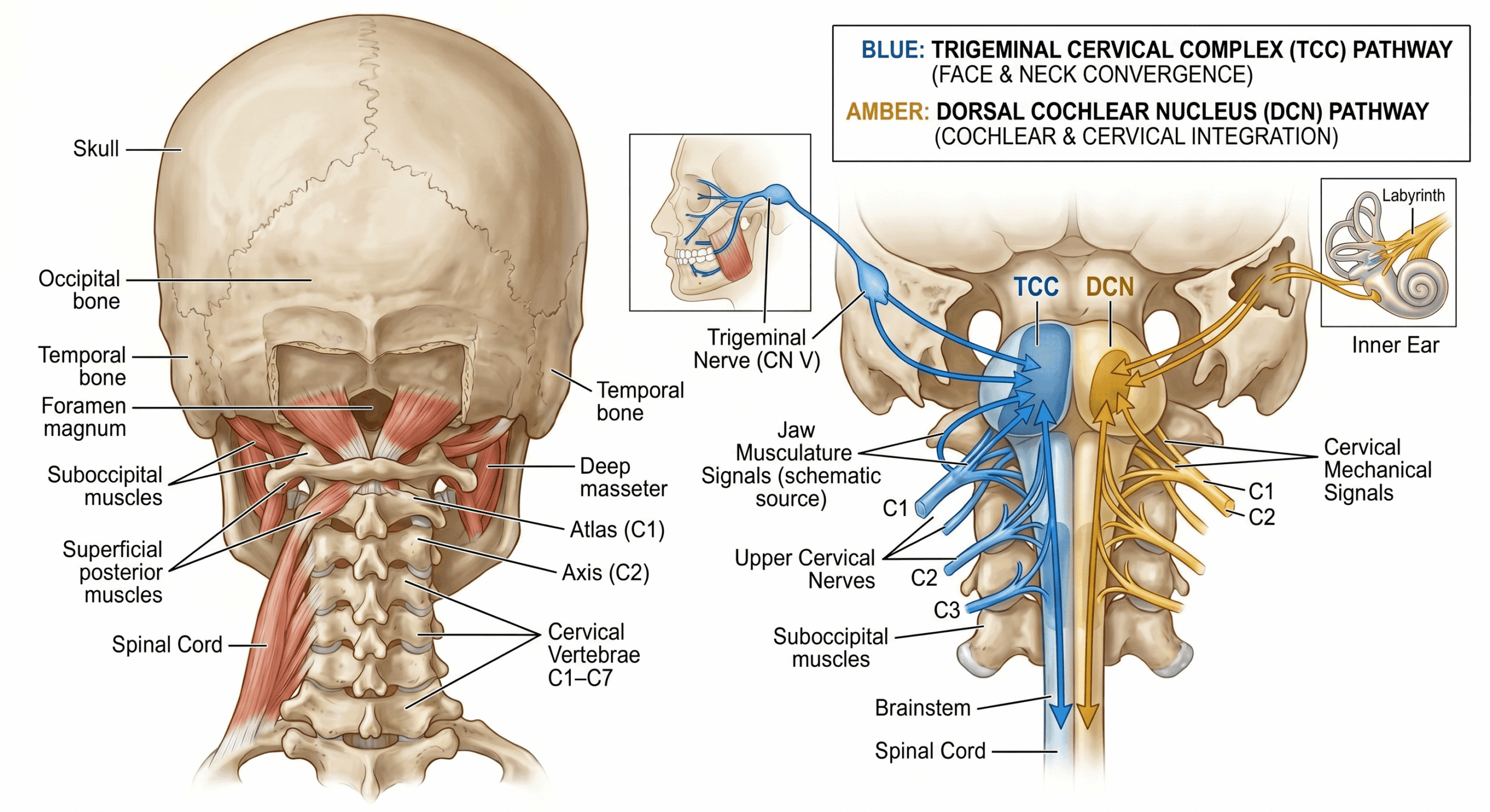
The anatomical basis for craniofacial referral from the neck is well documented. Nociceptive afferents from the upper three cervical nerves converge with afferents from the trigeminal nerve, which serves the face, jaw, and chewing muscles, in the trigeminocervical nucleus in the upper cervical spinal cord. Because of that convergence, pain arising from cervical structures can be referred into the face and head, and pain from the trigeminal territory can be referred back into the neck and occiput.
The NMF Science proposal is that chronic cervical strain contributes to sensitization at this junction, lowering the pain threshold across the craniofacial network, so that a flare of cervical tension can present as pain behind the eyes, jaw locking, or severe headache with the jaw as the endpoint rather than the origin. The convergence is established anatomy. The sensitization account of how cervical injury drives it is a hypothesis.
The auditory connection sits nearby. The dorsal cochlear nucleus, an auditory relay station in the brainstem, receives somatosensory input from the neck and jaw alongside signals from the ear, and this is the generally accepted anatomical explanation for why somatic modulation of tinnitus is possible at all. Exactly how cervical dysfunction translates into perceived sound is not settled, and accounts that describe the brain misreading mechanical pressure as sound go further than the evidence supports.
Does Treating the Neck Help the Tinnitus?
This is the question that matters, and there is trial evidence, though it is early and the studies are small.
A randomized controlled trial applied kinesio taping to the sternocleidomastoid, upper trapezius, and levator scapulae muscles in 30 patients with somatosensory tinnitus associated with neck complaints, against a sham taping control. Tinnitus severity, tinnitus handicap, cervical pain, and neck disability all improved significantly in the taping group. The sham group showed no significant difference at four weeks.
A second randomized trial in 40 patients with cervicogenic somatosensory tinnitus found that adding a supervised physical therapy exercise program to photobiomodulation therapy produced significantly greater improvement in tinnitus visual analog scores, tinnitus handicap inventory scores, and cervical range of motion than photobiomodulation alone.
Thirty and forty participants are small numbers, and neither trial tested the neuromyofascial approach specifically. What they establish is narrower and still useful: in patients whose tinnitus has a somatic component, physical intervention directed at cervical musculature can measurably change the tinnitus. That is not a claim anyone could have made from mechanism alone.
Why TMJ Is More Than a Jaw Problem
Temporomandibular joint dysfunction is conventionally understood as a mechanical problem with the jaw joint. Clicking, locking, and pain are attributed to joint misalignment, dental occlusion, or stress-related clenching, and treatment follows: bite guards, dental adjustment, local physiotherapy, sometimes surgery on the joint.
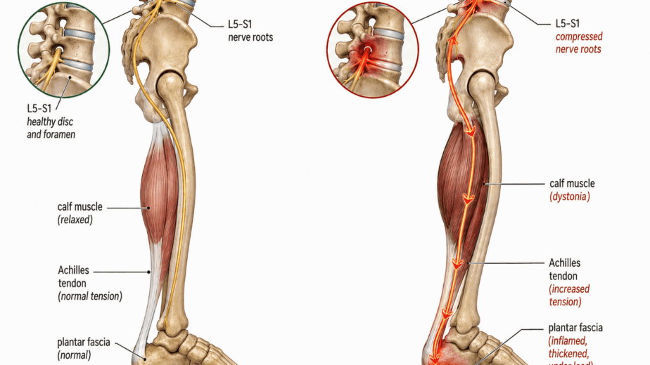
In my clinical experience, many TMJ presentations involve dystonia in the jaw muscles, meaning chronic involuntary spasm driven by abnormal signaling reaching the jaw rather than by a problem within the joint. When I map injury patterns in patients presenting with significant TMJ dysfunction, I frequently find findings in the cervical spine, upper thoracic spine, and craniofacial soft tissue rather than in the joint itself, often following motor vehicle accidents, sports impacts, or accumulated postural strain. The temporalis, masseter, and pterygoid muscles respond with sustained spasm that pulls the jaw off its normal hinge mechanics.
That is a clinical observation rather than a demonstrated mechanism, and the honest version of the claim is narrower than “the jaw is never the problem.” Local jaw pathology is real and common. The proposal is that in a subset of patients, the joint is an endpoint and the driver sits higher up.
Jaw Grinding as a Clinical Signal
Bruxism appears in the validated somatosensory tinnitus screening criteria for a reason, and further research supports the connection. A study of oral behaviors found that for each additional point scored on the Oral Behaviors Checklist, there was an 8.2 percent greater chance of having somatosensory tinnitus. The same study found somatosensory tinnitus associated with dizziness and with neck and temporomandibular joint pain, and found that tinnitus modulation through somatic manoeuvres and palpation of masticatory and cervical muscles was significantly associated with the condition.
The authors are careful to note that bruxism is not exclusive to somatosensory tinnitus patients. Its frequency, rather than its presence, is what appears to matter.
In my practice, jaw grinding appearing alongside unexplained tinnitus, recurrent headache, chronic neck pain, a whiplash event, or a history of concussion or head and neck trauma is a reason to assess the cervical and upper thoracic spine thoroughly. Patients often arrive unaware of the grinding, having been told they may have sleep bruxism or having had a dental professional flag wear on their teeth, without anyone connecting it to the neck.
The Overlap with Other Conditions
Patients presenting with both tinnitus and TMJ dysfunction commonly carry other diagnoses as well, frequently post-concussion syndrome, chronic migraine, or fibromyalgia. Dizziness co-occurs often enough to appear in the research cited above.
From a standard medical model these are separate conditions managed by separate specialists. The neuromyofascial proposal is that in some patients they may represent different expressions of a shared injury pattern rather than unrelated diseases. That is a hypothesis requiring formal study, and it should not be read as a claim that one condition progresses into another or that these presentations share a confirmed cause.
What Assessment Should Cover
Standard audiology and dental assessment remain the appropriate starting points for tinnitus and TMJ dysfunction, and for many patients they are sufficient. Where symptoms persist despite appropriate local treatment, the cervical and upper thoracic spine deserve systematic assessment, and the somatic modulation test is a quick way to establish whether that is likely to be relevant.
The craniocervical junction at C0-C1 and C1-C2 is a frequent injury site in whiplash and concussive events, and given the trigeminocervical convergence described earlier, it is anatomically well placed to contribute to craniofacial and auditory symptoms. It is not routinely evaluated in standard tinnitus or TMJ workups.
The neuromyofascial approach maps the cervical and craniofacial findings present in a given patient and directs care accordingly rather than applying a standard protocol. Whether that produces better outcomes than existing cervical treatments for somatosensory tinnitus has not been tested in a controlled trial. The trials that exist suggest the general direction is worth pursuing.
For a meaningful group of patients, the jaw and the ear are where the symptoms are felt. The neck is worth examining before concluding that is where the problem lives.
The information in this article is educational and informational in nature. It is not intended as a substitute for professional medical advice, diagnosis, or treatment. If you are experiencing jaw pain, tinnitus, or related craniofacial symptoms, consult with a qualified healthcare provider to discuss the options appropriate for your situation.