When a patient describes waking up with a stiff neck, a migraine by noon, a numb hand by evening, and a familiar ache down the leg, the standard medical response routes each symptom through a different door. A neurologist for the head. An orthopedist for the hand. A pain specialist for the back. Each clinician assigns a label. Each label generates a treatment. And the patient returns home carrying five separate diagnoses, five separate explanations, and often, very little resolution.
I have spent more than thirty years examining that pattern, and I no longer believe those five symptoms are always separate problems.
The neuromyofascial science framework I developed is built around a different premise: that many of the most common and persistent pain presentations may be connected expressions of one underlying physical process. The symptoms look different because they surface in different parts of the body. The proposal is that the architecture producing them is often unified.
Understanding that architecture changes what you look for, and where.
What Is Acquired Neuromyofascial Pathology?
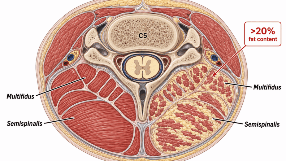
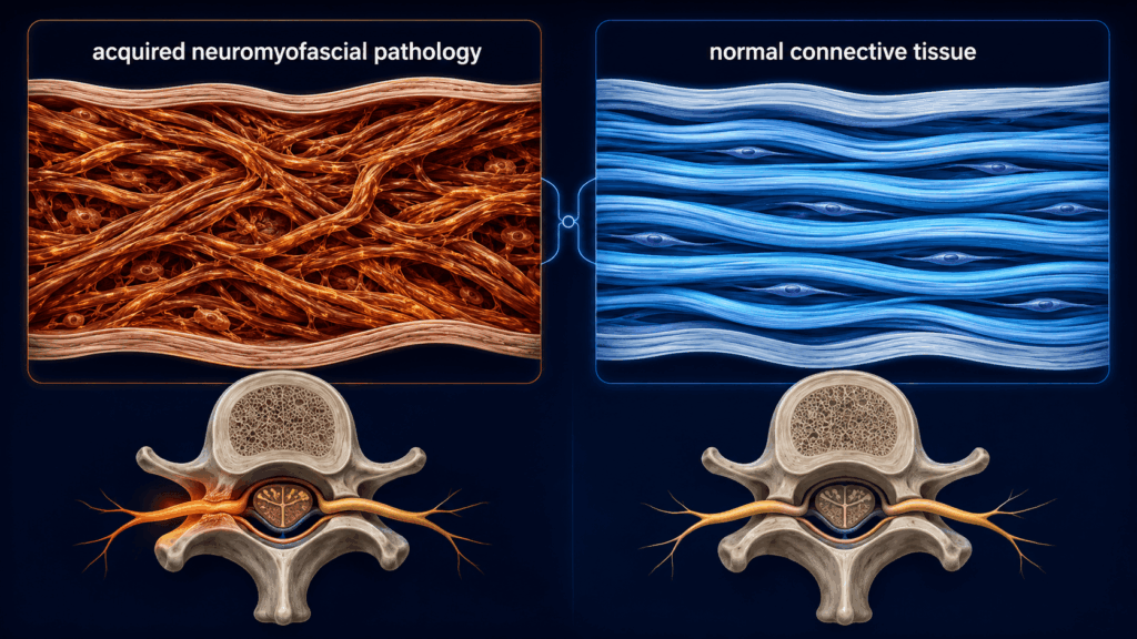
The central concept in this framework is what I refer to as acquired neuromyofascial pathology. This is not a single injury event. The proposal is that it is a cumulative process: over years and decades, microinjuries accumulate in predictable regions of the spine and limbs, scar tissue forms, and the density of the affected connective tissue increases. The working hypothesis is that this increased density begins to have mechanical consequences, including altered spinal positions, compressed joints, and irritation of the nerve roots passing through the region.
The process is slow, quiet, and difficult to see on standard imaging. Because the proposed damage lives in soft tissue density rather than in bone or disc, routine MRI and X-ray are poorly suited to detect it. Patients come in with real symptoms that do not correspond to findings on the scans used to look for them. The scan comes back clean, and the clinical response is often some variation of: this is just a normal part of getting older.
Fifty is still fifty. Sixty is still sixty. But the pain you are feeling is not always explained by age alone. In some cases, that pain may be the result of mechanical, structural burden that has been compounding for years.
Fascia Is Not Passive Wrapping
For a long time, the connective tissue scaffolding of the body, fascia, was treated as anatomically inert. It was considered wrapping. Background material. Anatomists dissected it away to reach the structures underneath.
That understanding has been substantially revised. Research reviewed by Gromakovskis (2025) supports the position that fascia is a richly innervated, biologically active tissue. It contains nociceptors, sympathetic fibers, and mechanoreceptors. When this tissue undergoes pathological change, including densification, fibrosis, and impaired sliding between tissue layers, it may not be a passive bystander to the pain process. That review describes fascia as a potential peripheral driver of myofascial pain, while noting that the current evidence remains preliminary and heterogeneous.
This matters clinically because it changes the target. If the connective tissue itself is pathological, treating only the downstream symptom may miss the source.
Measuring What Cannot Be Seen on MRI
One of the most useful recent developments in this area is the application of diagnostic ultrasound to connective tissue mechanics. Tomita and colleagues (2025) measured elevated thoracolumbar fascia shear strain in patients with nonspecific low back pain compared with asymptomatic individuals, in 32 patients and 32 controls. Those elevations correlated with pain and disability scores, while fascia thickness was comparable between groups.
The direction of that abnormality is still being worked out. Vining and colleagues (2023) describe shear strain between fascia layers as reduced in chronic low back pain rather than elevated. Two research groups, measuring the same tissue with similar technology, currently report opposite directions of change. That disagreement is worth stating plainly, because it tells you the field is early. What both groups agree on matters more than where they differ: the mechanical behaviour of fascia is measurably different in people with chronic back pain, and it can be quantified.
More recently, Tomita and colleagues (2026) ran a randomized trial testing whether those mechanics can be changed. Sixty participants received acupuncture, chiropractic care, or no treatment, with the untreated group later receiving massage. Chiropractic care reduced cumulated fascia shear strain by roughly 16 percent and massage by roughly 32 percent, while acupuncture produced no detectable change. In the untreated control period, shear strain increased. Only the chiropractic group showed improvement in disability scores.
One finding in that trial deserves emphasis because it cuts against a simple story: the change in fascia mechanics did not correlate with the change in disability. Manual therapy moved the tissue measurement and moved the clinical outcome, but not in lockstep. That is a caution against assuming that a mechanical measurement is the same thing as the patient’s experience, and it is precisely the kind of question that needs formal study rather than assumption.
What this body of work establishes is narrower than a vindication of any framework, and more useful. Fascial mechanics can be measured. They differ in people with chronic pain. They change in response to physical intervention, and they drift in the absence of it. The neuromyofascial framework proposes that those mechanics are a meaningful part of what produces chronic symptoms in some patients. The imaging work suggests that proposition is testable, which is the necessary first step toward knowing whether it is correct.
The Spine-to-Limb Chain
One of the clearest places to examine this connected architecture is carpal tunnel symptoms and hand numbness.
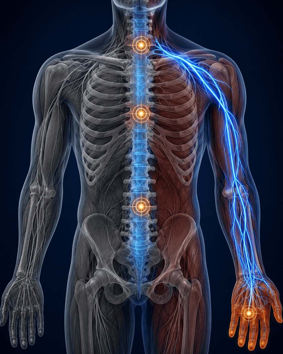
The intuitive assumption is that a numb hand is a wrist problem. That is where the symptoms are. That is where the standard diagnosis lands. But in the neuromyofascial framework, numbness and tingling in the hand may be downstream signals from a disruption further up the chain. The site of pathology may be in the neck, at the shoulder outlet, at the axilla, or at the elbow. The wrist may be a terminal expression of a restriction that originated above it.
The medical literature engages this logic through the concept of double crush syndrome, a recognized clinical framework in which concurrent cervical radiculopathy exists alongside a peripheral nerve entrapment such as carpal tunnel syndrome. The evidence on how best to treat it is still developing, and it is not one-sided. Hansen and colleagues (2024) found that patients with double crush syndrome improved after carpal tunnel release at rates comparable to patients with carpal tunnel syndrome alone, and concluded that carpal tunnel release is a reasonable first step before proceeding to cervical decompression.
Surgical sequencing data points in a different direction. Holloway and colleagues (2025) examined patients who underwent both cervical and peripheral nerve decompression and found that those who had cervical decompression first showed significantly better functional outcomes at one month, six months, and one year. Primary cervical decompression was associated with roughly 2.5 times the odds of achieving a clinically meaningful improvement in physical function. Patients who had both carpal and cubital tunnel release, rather than one alone, also did better. The authors concluded that cervical pathology may have a greater impact on overall outcomes and should be prioritized in surgical planning.
That is the clinical logic of the spine-to-limb chain: the proximal contribution appears to matter, and the sequence in which the pathway is addressed may affect the result. The same logic applies to presentations well beyond carpal tunnel.
Mapping the Architecture
If the standard examination and standard imaging are not designed to locate these injury sites, a different method is required.
A specialized neuromyofascial examination is a physical process. It relies on manual evaluation of the tissue itself, identifying regions of abnormal density, restricted sliding, and altered mechanics that do not produce findings on MRI. In more advanced cases, additional clinical confirmation can help establish the location and nature of the suspected pathology.
The objective of this process is not to assign a new diagnostic label. Labels are descriptions of symptoms. What the neuromyofascial audit produces is a map: where the tissue appears abnormal, how dense it is, and which nerves, joints, or spinal regions may be mechanically compromised as a result. That map informs the care pathway.
If findings are identified early, targeted self-care and tissue remodeling approaches may address the density before it compounds further. In more advanced cases, where decades of accumulation have produced significant structural burden, more intensive non-interventional or interventional approaches may be required. The map does not just identify what is present. It indicates how far the pattern has progressed, and what level of intervention the tissue may need.
Why This Matters for Patients Who Have Not Found Answers
The patients who spend years carrying multiple diagnoses, cycling through specialists, and completing treatment after treatment without sustained improvement are not failing to respond. In some cases, they may be being treated for the output while the input remains unaddressed.
Where a stiff neck, a migraine, a numb hand, morning stiffness, and sciatica trace back to a shared architecture of acquired soft tissue pathology, treating each symptom individually may be an incomplete strategy. The relief, when it comes, tends to be partial and temporary, because the compounding process continues if the source has not been found.
Neuromyofascial science is an attempt to answer a different question: not what label fits the symptom, but what physical site may be producing it. The embedded resources on this page, including a full explainer video and an annotated slide presentation, walk through the specific anatomy and clinical evidence in detail. The written summary above is the framework. The media is the mechanism.
If you want to understand what may be happening in your body, start there.
This article is written for educational purposes and represents the clinical perspective of Dr. G. Blair Lamb as developed through the neuromyofascial science framework. It is not intended as personal medical advice or as a substitute for individualized clinical evaluation. If you are experiencing chronic pain or neurological symptoms, consult a qualified healthcare provider.