One of the most frustrating experiences in medicine is a patient who has been in significant pain for months or years, has undergone every standard test, and keeps receiving the same answer: your imaging is normal.
The imaging is not lying. But it may not be telling the whole story.
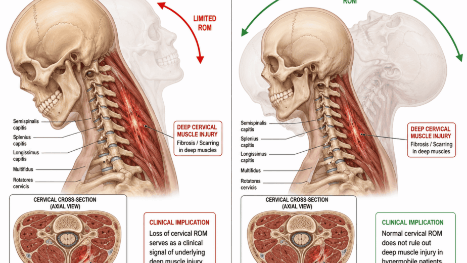
For nearly three decades I have been interested in what standard imaging is not designed to detect: structural change in deep spinal muscle tissue after injury. There is now a measurable body of research on this, and it is worth understanding both what it establishes and where it stops.
What Standard Imaging Can and Cannot See
X-ray, CT, and MRI are excellent tools for identifying fractures, disc herniations, gross anatomical abnormalities, and tumours. They are not designed to quantify changes within the spinal muscles themselves, particularly in the weeks and months following a whiplash event.
It is common to see patients who have been in a motor vehicle accident, who develop chronic spinal pain, and whose imaging reports come back normal or near normal. That does not establish that nothing happened to their spine. It establishes that nothing appeared on a study not built to look for this.
There have been advances in soft tissue spinal MRI and in spinal ultrasound over the past two decades. They are meaningful, and they remain limited in routine clinical practice.
Fat and Water Imaging: What It Measures
Fat and water MRI measures the proportion of fat within a muscle directly, rather than inferring it from appearance. Applied to the deep cervical muscles after injury, it has produced some of the most useful data in this field.
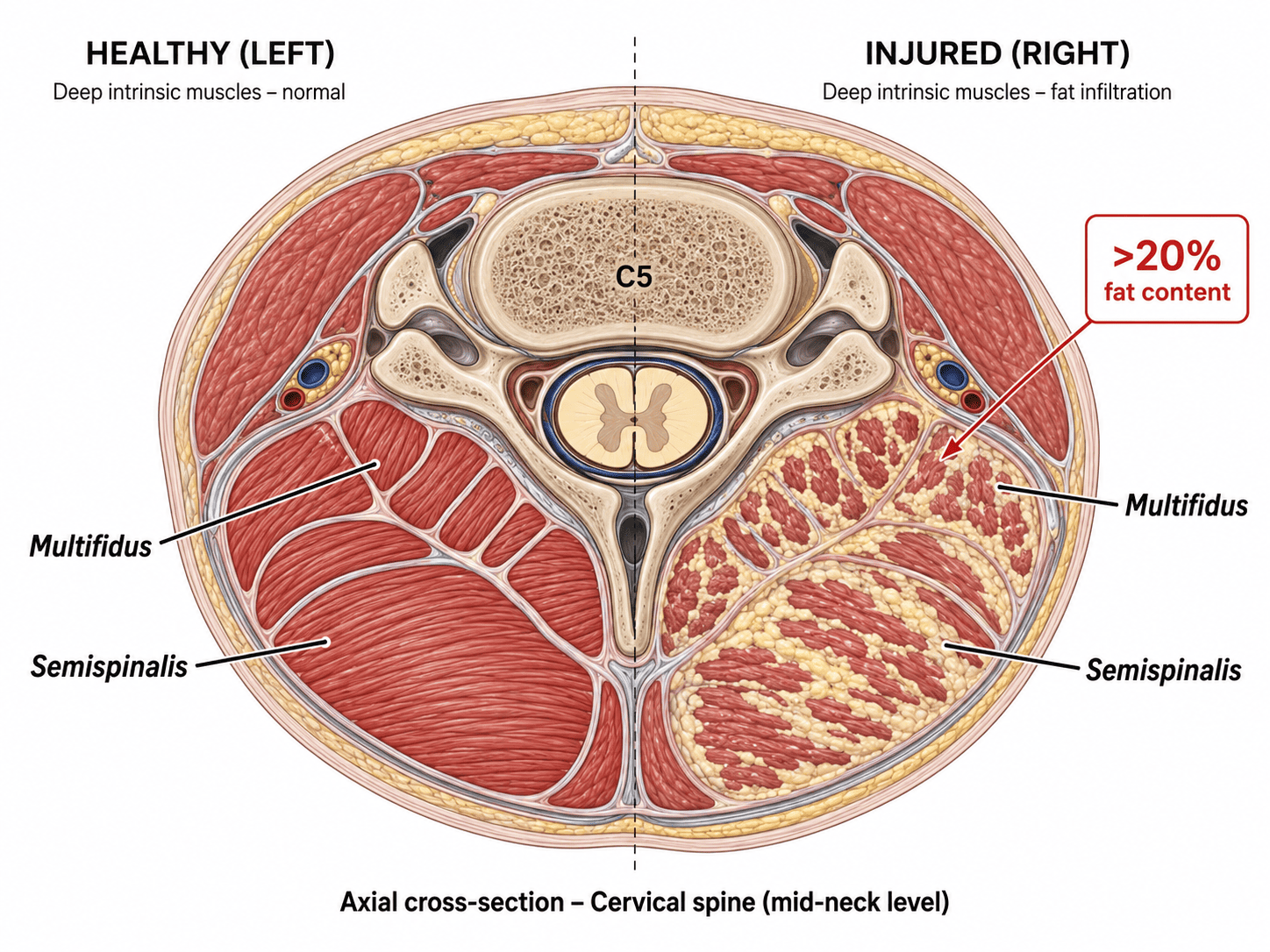
I describe the underlying process as spinal marbling, by analogy with a heavily marbled cut of beef. Functional contractile muscle is progressively displaced by fat. That is a descriptive term of mine rather than a clinical one, but the phenomenon it describes is measurable.
In a prospective study, 36 patients were imaged within one week of whiplash injury, again at two weeks, and at three months. There was no difference between groups at enrolment. By two weeks, patients who would go on to report severe disability at three months already showed significantly higher fat infiltration in the cervical multifidus than those who recovered. A threshold of 20.5 percent fat infiltration at two weeks predicted poor outcome at three months with 87.5 percent sensitivity and 92.9 percent specificity. The authors concluded that muscle degeneration occurs soon after injury, but only in patients with poor functional recovery, and that routine imaging protocols may need to be reconsidered.
A larger cohort of 97 patients followed to twelve months found that neck muscle fat infiltration at one week, together with a traumatic distress score, significantly predicted neck disability at one year. A confirmatory study found the infiltration concentrated in the medial portions of the deep cervical extensors, with the magnitude significantly greater in patients with severe chronic symptoms.
An earlier study of 44 patients found something that complicates a purely mechanical reading: initial post-traumatic stress symptom severity mediated the relationship between pain intensity and fat infiltration at six months, while loss of range of motion did not. The authors of the 97-patient study were likewise explicit that it is unclear whether higher fat infiltration represents a pre-existing characteristic or a result of the trauma.
So the picture is genuinely interesting and genuinely unfinished. The tissue change is real, measurable within two weeks, and strongly predictive of who does badly. How it arises, and whether it is cause or consequence, is not settled.
The Lower Back: A More Cautious Picture
Similar work has been done in the lumbar spine, and the findings there are more mixed. They are worth reporting accurately rather than selectively.
Mengiardi and colleagues used proton MR spectroscopy to measure fat content in the lumbar multifidus and longissimus muscles of 25 patients with chronic low back pain and 25 asymptomatic volunteers matched for age, sex, and body mass index. Mean fat content in the multifidus was 23.6 percent in the pain group and 14.5 percent in the volunteers, a significant difference. In the longissimus, the difference was not significant.
Two further findings deserve attention. Using a semiquantitative grading scale, the differences between patients and volunteers were not significant, and nearly half the pain patients graded zero for multifidus fat, as did just over half the volunteers. And no significant correlation was found between fat content and pain intensity, pain duration, or self-rated disability.
That is a more restrained result than the cervical whiplash data. Fat content in the lumbar multifidus is elevated as a group difference in chronic low back pain, but it does not track with how much pain an individual patient has or how long they have had it. Anyone citing this work as evidence that fat infiltration explains low back pain is citing more than it says.
The Shoulder: Where Nerve and Tendon Can Be Distinguished
The rotator cuff is where the mechanism behind fatty infiltration has been studied most directly, and the findings are relevant well beyond the shoulder.
Fatty infiltration of the supraspinatus is measured routinely in shoulder practice. It correlates with tear size, it is an independent risk factor for retear after surgical repair, and its severity predicts treatment outcome. The conventional explanation is that the tear comes first, with tendon retraction and reduced loading driving the fatty change that follows.
That explanation is not the whole picture. A controlled laboratory study separated the two mechanisms directly. Twenty-two rabbits underwent bilateral shoulder procedures randomized to suprascapular nerve transection alone, tendon transection alone, both combined, or control. By six weeks all groups showed muscle atrophy along with increasing adipocytes and fibrosis on histology. Between six and twelve weeks, only the isolated nerve injury group showed a continuing increase in fatty infiltration. The authors concluded that tendon injury and nerve injury are each independently associated with increased fatty infiltration and muscle fibrosis.
The distribution differed as well. Tendon injury concentrated fat near the myotendinous junction. Nerve-mediated fatty infiltration was diffuse.
That distinction holds in humans. A comparative analysis by Beeler, Ek and Gerber examined 20 shoulders with chronic rotator cuff tears and no electromyographic evidence of suprascapular neuropathy, against 17 shoulders with documented suprascapular nerve dysfunction and no tear. Muscle changes following chronic cuff tear differed from those following denervation, particularly in the appearance of the muscle border, the degree of perineural fat, and the overall distribution of infiltration. The authors concluded that highly specific and characteristic morphological patterns exist for each.
Two things follow, and they matter for the wider argument here. Nerve injury alone is sufficient to produce fatty infiltration in skeletal muscle, with no tendon damage at all. And the fat resulting from nerve injury is distinguishable on imaging from the fat resulting from mechanical injury, meaning the two causes are separable rather than interchangeable.
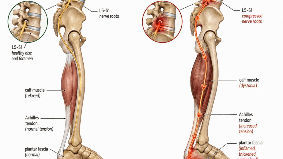
Whether nerve involvement originating in the cervical spine contributes to fatty change in shoulder or spinal musculature in the way I propose has not been studied. What this research establishes is that the mechanism is real in principle: compromise the nerve supply to a muscle and fat accumulates within it, in a pattern distinguishable from other causes. That is a foundation for the question rather than an answer to it.
Why This Matters Clinically
Fat and water imaging is not new in research. It has been accumulating in the literature for two decades. What it has not done is translate into routine clinical practice, and most patients presenting with chronic spinal pain after whiplash are assessed with protocols not designed to detect this type of change.
The clinical implication in the cervical whiplash setting is reasonably strong. A measurement taken at two weeks predicted three-month outcome with high sensitivity and specificity in the study cited above. If that finding replicates in larger cohorts, early identification could change how care is sequenced and how closely patients are followed.
It is worth being honest about what remains unknown. I am not aware of studies establishing whether deep cervical fat infiltration is reversible, or whether any intervention reduces it, or whether reducing it changes outcomes. The research shows it predicts who does badly. Whether it is a target or a marker is a separate question that has not been answered.
Much of the NMF Science investigational framework is built around intrinsic spinal pathology as a contributor to chronic pain. This imaging research provides measurable evidence that deep spinal tissue undergoes structural change after injury that standard imaging does not capture and that predicts poor recovery, and the shoulder work establishes that nerve compromise alone can produce that kind of change. It does not confirm the wider framework, and I would not present it as doing so. It confirms that something structural is happening early in the patients who do not recover, and that nerve involvement is a plausible route to it. Those are narrower claims and better supported ones.
Further research is needed, and I expect it to refine both the diagnostic thresholds and the clinical applications of these findings.
The information in this article is educational and informational in nature. It is not intended as a substitute for professional medical advice, diagnosis, or treatment. If you are experiencing chronic pain or have questions about your imaging results, consult with a qualified healthcare provider.