One of the most frustrating experiences in medicine is a patient who has been in significant pain for months or years, has undergone every standard test, and keeps receiving the same answer: your imaging is normal.
The imaging is not lying. But it is not telling the whole story either.
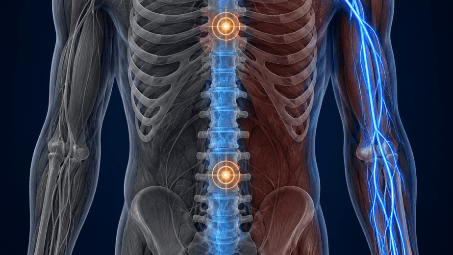
For nearly three decades, I have been studying what standard imaging consistently misses: the structural transformation of deep spinal muscle tissue after injury. Understanding this process is central to understanding why so many patients with chronic spinal pain do not recover with conventional rehabilitation, and what can be done about it.
What Standard Imaging Can and Cannot See
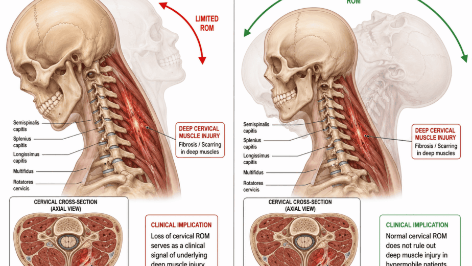
X-ray, CT scan, and MRI are excellent tools for identifying fractures, disc herniations, gross anatomical abnormalities, and tumors. They are not well designed to detect changes in the soft tissue around and within the spinal muscles, particularly in the weeks and months following a whiplash event.
It is common to see patients who have been in a motor vehicle accident, who develop chronic spinal pain, and whose imaging reports come back as normal or near-normal. This does not mean nothing happened to their spine. It means the injury occurred in tissues that standard protocols are not tuned to see.
There have been advances in soft tissue spinal MRI over the past decade, and similar progress in spinal ultrasound. These developments are meaningful, but they remain limited in clinical practice.
Fat Water Indexing and Spinal Marbling
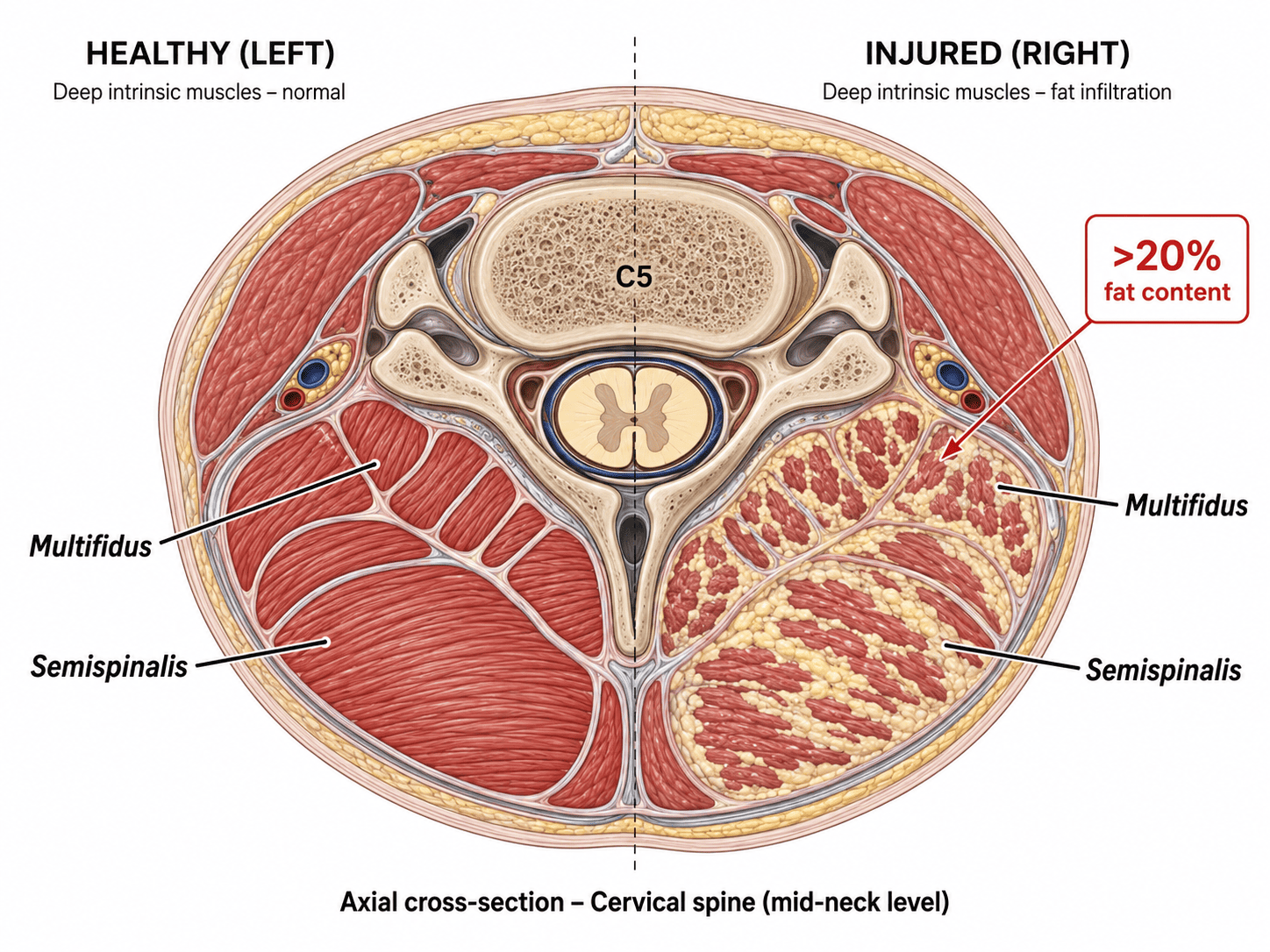
The MRI analysis technique called fat water indexing offers a more informative look at what happens to spinal muscle tissue after injury. The principle is straightforward: following trauma, deep spinal muscles can become injured and progressively replaced by fat tissue over time. This fat infiltration typically begins developing around three months after a whiplash event and continues as the damaged muscle is replaced by scarred, fatty tissue.
I describe this process as spinal marbling, a reference to what you see in a cut of heavily marbled beef. The muscle tissue, rather than remaining functional and contractile, is progressively displaced by fat. You cannot see this on a standard MRI report, but using fat water indexing, the fat content within the muscle can be measured directly.
Researcher James Elliott and his colleagues have demonstrated this process repeatedly in cervical spine studies following whiplash injuries. Their findings showed that fat infiltrates begin forming as early as two weeks after a motor vehicle accident. At the two-to-four week mark, the degree of fat infiltration in the cervical muscles could predict, with meaningful accuracy, which patients would recover with standard rehabilitation and which would not. By three months, the fat marbling in the deep spinal muscles was clearly visible and directly associated with chronic pain, failure to recover, and in some cases, the development of anxiety and PTSD symptoms.
Across multiple studies using fat water indexing MRI of the spine, a consistent finding emerges: when fat content in the deep spinal muscles exceeds approximately 20 percent, persistent pain is likely and standard rehabilitation is unlikely to produce full recovery.
The Same Pattern in the Lower Back and Shoulder
This process is not limited to the neck. Research groups led by Mengiardi demonstrated similar fat infiltration patterns in the lower back, where the intrinsic spinal muscles showed higher concentrations of fat in patients with chronic low back pain compared to asymptomatic volunteers.
Notably, fat infiltration was present in all chronic low back pain patients in that study, whether the pain originated from a specific injury event, repetitive strain, or aging-related wear. This suggests that intrinsic spinal muscle scarring and fat replacement may be a common pathway underlying most spinal pain problems, not just those following acute trauma.
The research group led by Pfirrmann extended this finding to the rotator cuff as early as 2004. In that work, fat content in the rotator cuff muscles was predictive of the degree of muscle tearing. Higher fat infiltration correlated with greater risk of tear. This finding pointed toward a sequence of events that fits closely with the neuromyofascial model: nerve signal loss from the cervical spine leads to denervation of the supraspinatus and surrounding rotator cuff muscles, which then shorten, scar, and accumulate fat, making them increasingly susceptible to tearing.
In other words, what looks like a shoulder problem may have its structural origin in the neck.
Why This Matters Clinically
Fat water indexing is not a new concept in research. It has been accumulating in the literature for over two decades. What it has not done is translate meaningfully into routine clinical practice. Most patients presenting with chronic spinal pain following whiplash are assessed with standard imaging protocols that were not designed to detect this type of tissue transformation.
The clinical implications of this are significant. A patient whose deep cervical muscles show greater than 20 percent fat infiltration at two to four weeks post-injury is unlikely to recover with standard physiotherapy alone. Early identification of this pattern could change the trajectory of care decisions, including the timing and type of interventions applied.
At NMF Science, much of our investigational framework is built around intrinsic spinal pathology as a primary driver of chronic pain. The fat water indexing research provides a measurable, reproducible confirmation of what clinical observation has indicated for years: that the deep soft tissue around the spine undergoes structural changes after injury that persist, progress, and generate chronic pain in ways that standard imaging cannot detect.
Further research in this area is needed, and I expect it will continue to refine both the diagnostic thresholds and the clinical applications of these findings.
The information in this article is educational and informational in nature. It is not intended as a substitute for professional medical advice, diagnosis, or treatment. If you are experiencing chronic pain or have questions about your imaging results, consult with a qualified healthcare provider.